Some big scientific discoveries aren’t actually discovered. They are borrowed. That’s what happened when scientists enlisted proteins from an unlikely lender: green algae.
Cells of the algal species Chlamydomonas reinhardtii are decorated with proteins that can sense light. That ability, first noticed in 2002, quickly caught the attention of brain scientists. A light-sensing protein promised the power to control neurons — the brain’s nerve cells — by providing a way to turn them on and off, in exactly the right place and time.
Nerve cells genetically engineered to produce the algal proteins become light-controlled puppets. A flash of light could induce a quiet neuron to fire off signals or force an active neuron to fall silent.
“This molecule is the light sensor that we needed,” says vision neuroscientist Zhuo-Hua Pan, who had been searching for a way to control vision cells in mice’s retinas.
The method enabled by these loaner proteins is now called optogenetics, for its combination of light (opto) and genes. In less than two decades, optogenetics has led to big insights into how memories are stored, what creates perceptions and what goes wrong in the brain during depression and addiction.
To celebrate our upcoming 100th anniversary, we’re launching a series that highlights some of the biggest advances in science over the last century. For more on the past, present and future of neuroscience, visit Century of Science: Our brains, our futures.
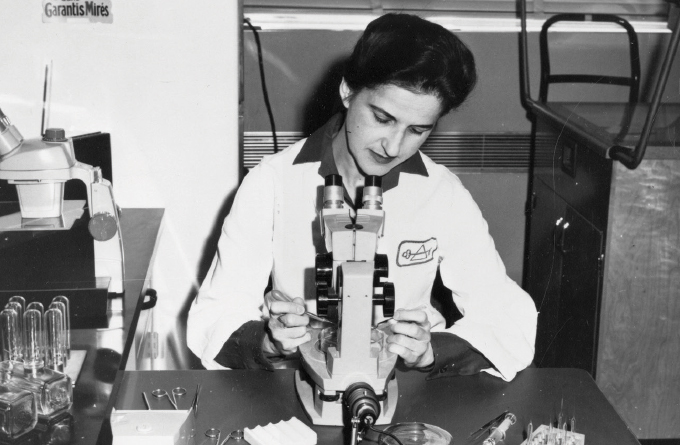
An early clue to the potential of optogenetics came around 1 a.m. on August 4, 2004. Neuroscientist Ed Boyden was in a lab at Stanford, checking on a dish of neurons that possessed a gene for one of the algal light sensors, called channelrhodopsin-2. Boyden was going to flash blue light on the cells and see if they fired signals. To his amazement, the very first cell he checked responded to the light with a burst of action, Boyden wrote in a 2011 account. The possibilities raised by that little spark of activity, described in a 2005 technical report by Boyden, Karl Deisseroth of Stanford University and colleagues, quickly became realities.
In Pan’s lab, light-responsive proteins restored vision in mice with damaged retinas, a finding that has now led to a clinical trial in people. Optogenetics’ promise wasn’t a given in those early days, as scientists were first learning how to use these proteins in neurons. “At that time, no one anticipated that this optogenetic work would have such a huge impact,” Pan says.
Since those early discoveries, the algae’s light sensors have been adopted for use in numerous brain research arenas. Neuroscientist Talia Lerner of Northwestern University in Chicago, for example, uses optogenetics to study connections between cells in the mouse brain. The method allows her to tease apart the relationships between cells that produce and respond to dopamine, a chemical messenger involved in movement and reward. Those cellular links, illuminated by optogenetics, might help reveal details about motivation and learning. “My research really wouldn’t be possible in its current form without optogenetics,” she says.
Optogenetics is also indispensable for Jeanne Paz of the Gladstone Institutes in San Francisco. She and her colleagues have been hunting for the cells that can stop seizures from spreading across the brain. By giving her a way to control distinct groups of neurons, optogenetics is crucial to her search. “We really could not ask these questions with any other tool,” Paz says.
Her optogenetics-aided search led Paz to a brain structure called the thalamus, a way station for many neural networks in the brain. “I remember the goose bumps I experienced the first time I shined the light into the thalamus and it stopped the seizure,” she says.
So far, optogenetics research has taken place mostly in mice. But insights into more complex brains, including those of primates, may soon be found, says Yasmine El-Shamayleh of Columbia University. In 2009, Boyden and colleagues described optogenetics in a macaque. El-Shamayleh and others are pushing this line of research, hard. “We are definitely on the cusp” of revealing some fascinating principles of the primate brain, such as how the brain transforms signals from the eyes into perceptions, she says.
Optogenetics has evolved quickly. Scientists have engineered and optimized new light sensors and new ways of combining them with other techniques. An important reason for today’s widespread innovation, says Lerner, was the early spirit of sharing by optogenetics pioneers. At Stanford, Deisseroth would regularly run workshops to train other scientists on the technique. “In some ways, that’s as important as inventing it,” Lerner says.
So it’s worth taking a minute to appreciate the original sharers. No matter what happens next in this swiftly moving field, one thing is certain: Brain scientists will be forever in the algae’s debt.
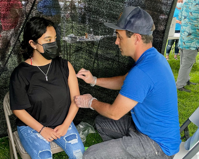
PHILADELPHIA — In a makeshift tent behind a soccer goal and close enough to a taco stand that the smell of grilling barbacoa and carnitas drifts over, Melissa Pluguez cheerfully asks a man, in Spanish, if he’s right- or left-handed.
The man, wearing jeans and a red T-shirt with white letters that spell Abercrombie, answers right, and confesses he’s a bit afraid of needles. Even so, he’s been eager to get a COVID-19 vaccine, Pluguez translates, but hasn’t felt comfortable going to vaccination sites around Philadelphia. But when he heard about this vaccination event — staffed by local, Spanish-speaking medical professionals and held at his church congregation’s regular Sunday gathering — he felt ready.
Pluguez is an emergency room nurse at Cooper University Health Care in Camden, N.J., and co-medical director of Unidos Contra COVID, the small group that organized this vaccine outreach event. She tells the man that the fear is worse than the needle, and he nods and looks away as she injects the Pfizer vaccine into his left arm. Afterward, he smiles, and the two bump elbows before the man leaves to pick up his vaccination card.
There’s no free beer in sight, nor is anyone getting complimentary tickets to Phillies baseball games. Instead, roughly 300 people are clustered around soccer fields that border the church parking lot. The main event is a tournament, where professional-looking players in uniform square off as spectators cheer. On adjacent fields, children kick balls around or chase each other through the lines of people waiting to buy tacos or mango slices stuffed into plastic cups. Couples dance to upbeat music emanating from loudspeakers set up near Unidos Contra COVID’s tent. Inside, behind dark mesh netting partitions set up for privacy amid all that action, vaccines are being delivered into arm after arm.
In the end, this sort of targeted approach may be a more impactful way than the flashier, broader efforts, like beer giveaways and large cash prizes, to reach the roughly 35 percent of adults in the United States who haven’t yet gotten at least one COVID-19 vaccine dose. Such a hyperlocal focus, experts say, can address the idiosyncratic and complex array of reasons that people aren’t getting vaccinated, especially in often overlooked and underserved communities that have experienced higher rates of COVID-19 and relatively low numbers of vaccinations (SN: 5/3/21).
Like in other parts of the country, Hispanic communities in Philadelphia have been especially impacted by COVID-19. Throughout the pandemic, hospitalization rates for Hispanic people in the city who are 35 and older have been higher than for any other group, as have death rates for those 75 and older. And vaccinations have lagged; only 37 percent of the Hispanic population has received one dose, compared with 51 percent of white residents as of June 14.
A group of local doctors and nursed formed Unidos Contra COVID, which means United Against COVID, earlier this year to address these disparities. The Hispanic population in Philadelphia is diverse, and “our experiences and makeup is hardly monolithic,” says José Torradas, an emergency room physician and co-medical director of Unidos Contra COVID, who left his job earlier this year to focus on outreach full-time. People in this community aren’t getting the vaccine “for different reasons in different groups.”
Figuring out those reasons has become crucial to Unidos Contra COVID’s mission. The group has found that for Central American and Mexican communities in parts of Philadelphia, access has been the biggest problem. Meanwhile, in the predominantly Puerto Rican and Dominican communities of northern Philadelphia, Torradas says, vaccine skepticism stems from misinformation and a general distrust of the government.
José Torradas, a physician and co-medical director of Unidos Contra COVID, rubs alcohol on a woman’s arm before giving her a COVID-19 vaccine shot at a local community gathering on June 13 in the Philadelphia area.Paula Lopez
Barriers to access
At the church event, Unidos Contra COVID came to bring vaccines to people who won’t travel to unfamiliar places to get them, because of language barriers or for fear of deportation. Many of the people gathered around the soccer fields are undocumented, Torradas says, though his group never asks about immigration status.
These Sunday gatherings represent the few hours each week that these communities come together, he says, often after church. “It’s a sanctuary, a place they feel safe.” For that reason, organizers asked that the church not be named.
The rest of the week, it’s either work or home for many undocumented people. “Anything outside of that routine represents a risk,” Torradas says. Even though local pharmacies may be close, or federal distribution sites accessible by bus, fears of interacting with government services or law enforcement keep many from getting vaccinated, he says. By law, undocumented immigrants are eligible to receive COVID-19 vaccines.
“There’s lots of desire [here] for the vaccine,” Torradas says. “They just don’t want to get deported.”
A Kaiser Family Foundation poll in May found that Hispanics who have yet to receive a COVID-19 vaccine are about twice as likely as non-Hispanic whites or Blacks to say they’d like to get vaccinated as soon as possible.
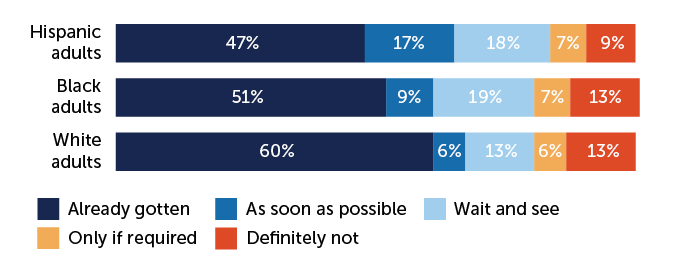
Shot status
Hispanic adults expressed a desire to get the COVID-19 vaccine “as soon as possible” at much higher rates than Black or white adults, according to a telephone survey of 2,097 adults conducted April 15 to April 29 by the Kaiser Family Foundation.
Intent to get a COVID-19 vaccine, by racial group
C. Chang
C. Chang
KFF COVID-19 Vaccine Monitor
To best serve these communities, Unidos Contra COVID teams up with trusted figures in the area, like José Hernández. He’s a church leader who can identify times and places where the group could have the most impact, and who can spread the word among the community.
Speaking loudly to be heard over the music, Hernández says that he had been trying to connect members of the congregation with vaccines since they first became available. But most people weren’t willing to travel far to get the shots, even though they are free. And simply going to their doctor often isn’t an option, since Hispanic Americans also have the highest rates of uninsurance of any racial or ethnic group according to the U.S. Census Bureau.
Being able to get the vaccines at a regular community event, and from people who speak Spanish, “has just been incredible,” Hernández says. Besides two large events that Unidos Contra COVID has held at his church, Torradas and Pluguez have both come out to give shots to smaller groups.
“We’ve never had an experience like this … where doctors come to us,” Hernández says.
Fear of deportation and language issues aren’t the only barriers to vaccination in some of these communities. There’s also gun violence to worry about. In 2020, there were over 2,200 shootings in Philadelphia, among the highest number for any U.S. city. Walking or taking the bus even just a kilometer or two to a vaccine distribution site in some neighborhoods “might mean crossing two or three gang turfs,” Torradas says. Some people have told him they don’t feel comfortable leaving their own block.
To reach such communities, Torradas and his colleagues have set up shop in places like schools and shopping malls, operating on weekends and evenings, in addition to church events. Many of the people who come “are day laborers who leave [home] at 5 a.m. and don’t get back till late,” Pluguez says. And many “are not hesitant. They are just not able to access the resources.”
Addressing vaccine hesitancy
Still, there are people in these communities who are skeptical of vaccines, Pluguez says. Reasons range from general distrust of government, to fears that stem from misinformation, such as that the vaccine causes sterility.
Some of the concern “is truly borne of crimes that have been committed [against] both Black and Hispanic communities,” Pluguez says, citing official programs that resulted in up to a third of Puerto Rican women being sterilized from the 1930s to 1970s, often without informed consent. “So hearing now that this vaccine might make them sterile? That runs deep, it runs very deep.”
Another common, false rumor is that the vaccines contain microchips with location trackers. Such rumors, which spread quickly on social media, can easily take hold in communities with undocumented immigrants. “They don’t want to have to fear every day that something they just put in their bodies is going to make them prone to being deported,” Pluguez says.
To address that mistrust, Unidos Contra COVID tries to identify trusted members of the community who could act as effective messengers. People are more likely to listen “to a face they recognize, who has been around longer than the vaccine,” Torradas says.
Individuals personally impacted by COVID, especially younger people, are especially effective. “Bring me the most fit person in this congregation who almost died of COVID and have him and the medical professionals stand in front of everybody, tell their story and answer their questions. That’s the formula for hyperlocal buy in,” he says.
Unidos Contra COVID also canvasses nearby streets during their vaccination events, trying to meet people where they are. “The moment I start speaking Spanish, you can absolutely see their eyes light up, and they start engaging in a way that’s very different,” Pluguez says. From that kernel of trust, she listens to people’s concerns about vaccination “without putting blame on them,” she says.
When misinformation comes up, she addresses it practically. “[If people ask] ‘What if there’s a microchip?’ I say, ‘Come look at the vaccine for yourself. Look at the needle. Look at the vial. You can see that there is nothing in it,’” Pluguez says. Often she talks about her experience as an emergency room nurse during the worst of the pandemic. “I share with them how many hands I’ve held of the sick and dying, alone,” she says, and how the vaccine can prevent that from happening.
At a previous vaccine outreach event in south Philadelphia, Pluguez once spent 45 minutes talking with one man she encountered walking down the street with his pregnant wife and three young boys. “He’d heard a lot of the rumors,” she says, about microchips and sterility. Pluguez addressed those concerns in Spanish, while playing with his kids to keep them entertained so she and the man could talk more. Twice during their conversation, the man left, saying he’d think more about it. “He made it a block before coming back with more questions,” she says.
Pluguez pulled up a chair for him, and they continued talking. As the conversation went on, she noticed a shift in his demeanor. “He started looking directly at me and making jokes,” she says. “That’s when I got the feeling that he finally trusts me.”
Finally, the man expressed worry about missing work if he got sick from the vaccine, Pluguez remembers. So she asked him, “What if you get sick with COVID? What is your wife going to do if you’re laid up in the hospital? Who is going to pay the bills?” Ultimately, that message got through.
“Then he said, ‘As long as you stand by my side, I’ll do it.’ And I said, ‘I’ll be here with you every step of the way,’” she says. Pluguez stayed with him through vaccine registration, the shot and the 15-minute waiting period afterward.
“I was really proud of him … I had such joy in my heart,” Pluguez says.
“We’re no longer looking at the people who were desperate to get vaccinated,” she says. “Now every single person who shows up is a victory, is one step further into making COVID an issue of the past.”
Amid the food, music and soccer at that Sunday vaccination event, 151 people got their first or second vaccine dose. Since Unidos Contra COVID began their events in early May, the group has vaccinated about 850 people, and hopes to scale up.
Toward the end of the afternoon event, Pluguez pulled out thick UPS package. “Our official 501(c)(3) documents,” she says. “I promised José I wouldn’t open them until we could do it together.”
They expect to get approval from the Internal Revenue Service in a few weeks. Becoming an official nonprofit will help the group more easily apply directly for grant money, allowing it to purchase more freezers for vaccine storage and hire more full-time staff. Currently, about 50 people contribute to the effort as volunteers, with Pluguez and Torradas devoting the most time.
The two pose for a picture with the documents, celebrating the milestone. “I may shed a tear, I’m so excited,” she says. Then, Pluguez gathers her things to leave for the hospital. Before she goes, someone from the taco stand brings her a Styrofoam box filled with dinner.
“I’ve worked the past six nights; tonight is my last shift,” she says. “I get off at 7 a.m., then I can sleep.”
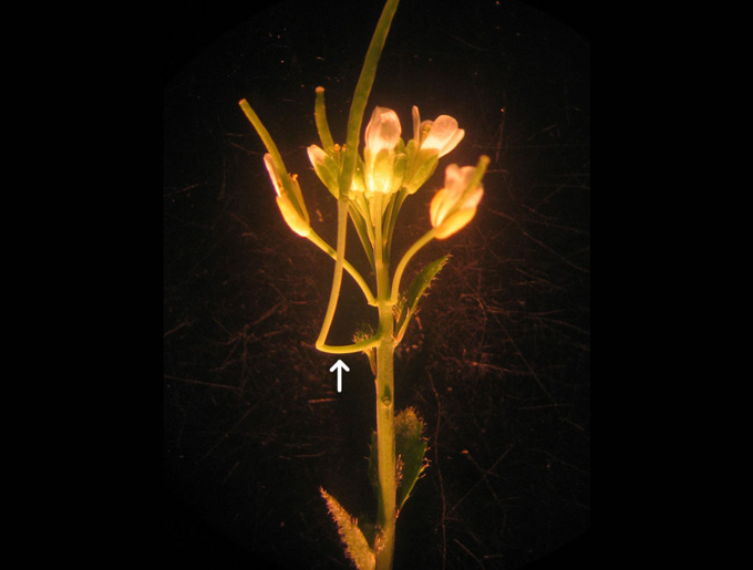
A common lab plant that’s been poked and put under microscopes for decades may seem unlikely to keep secrets. But in widely studied Arabidopsis thaliana, scientists have identified the “cantil” — a newly reported plant organ named for its cantilever-like way of branching off of the main stem. The structure appears in only some A. thaliana and only under certain conditions, researchers report online June 15 in Development.
“If you told me of a new organ in a weird plant in Amazonia, I wouldn’t be surprised at all,” says François Parcy, a plant biologist at CNRS in Paris, who was not involved in the study. “What struck me is this happened in Arabidopsis. This is something that’s really surprising.”
Molecular biologist Timothy Gookin first suspected contamination or a mutation when he noticed some A. thaliana with odd stalks jutting out from the stem, like half-finished bridges. It took 12 years of experiments at Penn State to show that the rare stalks are a new type of plant part and to explain their trigger: delayed flowering.
Like many other plants, short days prompt A. thaliana, which is in the same family as cabbage and mustard greens, to shore up resources; long days tell it to churn out flowers. Cantils form when that transition from stockpiling to blooming is postponed, as the plant keeps growing while waiting for the flowering signal, the researchers found. The cantil is “just growing kind of like, ‘Hey, where’s the summer? OK, I’m waiting for my break. Where’s it coming?’” Gookin says.
A newly described cantilever-like organ (arrow) depends on both genetics and circumstance to appear. It develops only in some Arabidopsis thaliana varieties, and only when flowering is postponed.T. Gookin
Scientists’ preference for using long growing days and fast-flowering conditions have helped keep cantils hidden in hundreds of labs worldwide, Gookin says.The organs can develop in the wild, though some favorite, fast-growing A. thaliana varieties have lost the genetic ability to produce cantils. It’s unclear how the plants use the organs.
Cantils are, so far, known to occur only in A. thaliana. So the plant parts may not rewrite biology textbooks just yet. But after being found in a lab plant that’s scrutinized so widely, it’s a reminder to keep observing closely (SN: 10/22/18).
Despite their mysterious nature, black holes are thought to follow certain simple rules. Now, one of the most famous black hole laws, predicted by physicist Stephen Hawking, has been confirmed with gravitational waves.
According to the black hole area theorem, developed by Hawking in the early 1970s, black holes can’t decrease in surface area over time. The area theorem fascinates physicists because it mirrors a well-known physics rule that disorder, or entropy, can’t decrease over time. Instead, entropy consistently increases (SN: 7/10/15).
That’s “an exciting hint that black hole areas are something fundamental and important,” says astrophysicist Will Farr of Stony Brook University in New York and the Flatiron Institute in New York City.
The surface area of a lone black hole won’t change — after all, nothing can escape from within. However, if you throw something into a black hole, it will gain more mass, increasing its surface area. But the incoming object could also make the black hole spin, which decreases the surface area. The area law says that the increase in surface area due to additional mass will always outweigh the decrease in surface area due to added spin.
To test this area rule, MIT astrophysicist Maximiliano Isi, Farr and others used ripples in spacetime stirred up by two black holes that spiraled inward and merged into one bigger black hole. A black hole’s surface area is defined by its event horizon — the boundary from within which it’s impossible to escape. According to the area theorem, the area of the newly formed black hole’s event horizon should be at least as big as the areas of the event horizons of the two original black holes combined.
The team analyzed data from the first gravitational waves ever spotted, which were detected by the Advanced Laser Interferometer Gravitational-Wave Observatory, LIGO, in 2015 (SN: 2/11/16). The researchers split the gravitational wave data into two time segments, before and after the merger, and calculated the surface areas of the black holes in each period. The surface area of the newly formed black hole was greater than that of the two initial black holes combined, upholding the area law with a 95 percent confidence level, the team reports in a paper to appear in Physical Review Letters.
“It’s the first time that we can put a number on this,” Isi says.
The area theorem is a result of the general theory of relativity, which describes the physics of black holes and gravitational waves. Previous analyses of gravitational waves have agreed with predictions of general relativity, and thus already hinted that the area law can’t be wildly off. But the new study “is a more explicit confirmation,” of the area law, says physicist Cecilia Chirenti of the University of Maryland in College Park, who was not involved with the research.
So far, general relativity describes black holes well. But scientists don’t fully understand what happens where general relativity — which typically applies to large objects like black holes — meets quantum mechanics, which describes small stuff like atoms and subatomic particles. In that quantum realm, strange things can happen.
For example, black holes can release a faint mist of particles called Hawking radiation, another idea developed by Hawking in the 1970s. That effect could allow black holes to shrink, violating the area law, but only over extremely long periods of time, so it wouldn’t have affected the relatively quick merger of black holes that LIGO saw.
Physicists are looking for an improved theory that will combine the two disciplines into one new, improved theory of quantum gravity. Any failure of black holes to abide by the rules of general relativity could point physicists in the right direction to find that new theory.
So physicists tend to be grumpy about the enduring success of general relativity, Farr says. “We’re like, ‘aw, it was right again.’”
Earth is on an orderly path around the sun, orbiting in nearly the same plane as our star’s equator. In 2008, however, astronomers began finding worlds in other solar systems that sail far above and below their star’s equatorial plane.
Now a surprising discovery about these wrong-way worlds may eventually reveal their origin: Most of them follow polar orbits (SN: 6/17/16). If Earth had such an orbit, every year we’d pass over the sun’s north pole, dive through its equatorial plane, then pass below the sun’s south pole before coming back up again.
Astronomers Simon Albrecht and Marcus Marcussen at Aarhus University in Denmark and colleagues analyzed 57 planets in other solar systems for which the researchers could determine the true tilt between a planet’s orbit and its star’s equatorial plane. Two-thirds of the planets have normal orbits, tilted no more than 40 degrees, the team found. The other 19 planets are misaligned.
But the orbits of those misaligned planets don’t make just any old angle with their star’s equator. Instead, they pile up around 90 degrees. In fact, all but one of the misaligned planets are on polar orbits, having tilts from 80 to 125 degrees, the astronomers report online May 20 at arXiv.org.
“It’s very, very strange,” says Amaury Triaud, an astronomer at the University of Birmingham in England who has found a number of misaligned planets but was not involved with the new study. “It’s a beautifully executed idea, and the result is most intriguing,” he says. “It’s so new and so weird.”
The result may lend insight into the biggest mystery about these planets: how they arose (SN: 10/18/13). Such worlds were a shock to astronomers, because planets form inside pancake-shaped disks of gas and dust orbiting in their stars’ equatorial planes. Thus, planets should lie near the plane of their sun’s equator, too. In our solar system, for example, Earth’s orbit tilts only 7 degrees from the solar equatorial plane, and even Pluto — which many astronomers no longer call a planet — has an orbit tilted only 12 degrees from that plane (and 17 degrees from the Earth’s orbital plane).
“At the moment, we are not sure what is the underlying mechanism” or mechanisms for creating misaligned planets, Albrecht admits. Whatever it is, though, it should account for the newly discovered plethora of perpendicular planets, he says.
A possible clue, Albrecht says, comes from the single exception to the rule: the one misaligned planet in the sample that is not on a polar orbit. This planet also happens to be the most massive in the sample, packing the mass of between five and eight Jupiters. Albrecht says that may be just a coincidence — or it may reveal something about how the other planets became misaligned.
In the future, the astronomers hope to understand how these wayward worlds acquired their odd orbits. All known misaligned planets orbit close to their stars, but are these worlds more likely than normal, close-in planets to have giant planets near them? The scientists don’t yet know, but if they find such a correlation, those companions may have somehow flung these bizarre worlds onto their peculiar planetary paths.
Researchers at UC San Diego, San Diego State University, and international collaborators have designed and validated a prediction model to signal counties at risk of future overdose death outbreaks.
There are only so many ways to cram DNA into a cell’s nucleus, a study suggests.
A cell’s complete genetic blueprint, or genome, is densely packed into chromosomes, condensing meters of DNA into a minuscule cellular vessel only micrometers wide (SN: 8/24/15). But how chromosomes fold to fit inside the nuclei of diverse species is unclear.
There appear to be two methods to stuff all of that DNA in, researchers report in the May 28 Science. Cells can even flip-flop which arrangement they have by inactivating a molecule called condensin II, the team found.
If chromosomes were pieces of paper, some, like those of humans, would look like a crumpled ball inside the nucleus, says Claire Hoencamp, a molecular biologist at the Netherlands Cancer Institute in Amsterdam (SN: 10/8/09). Others, like those of fruit flies (Drosophila melanogaster), resemble flat sheets of stacked paper.
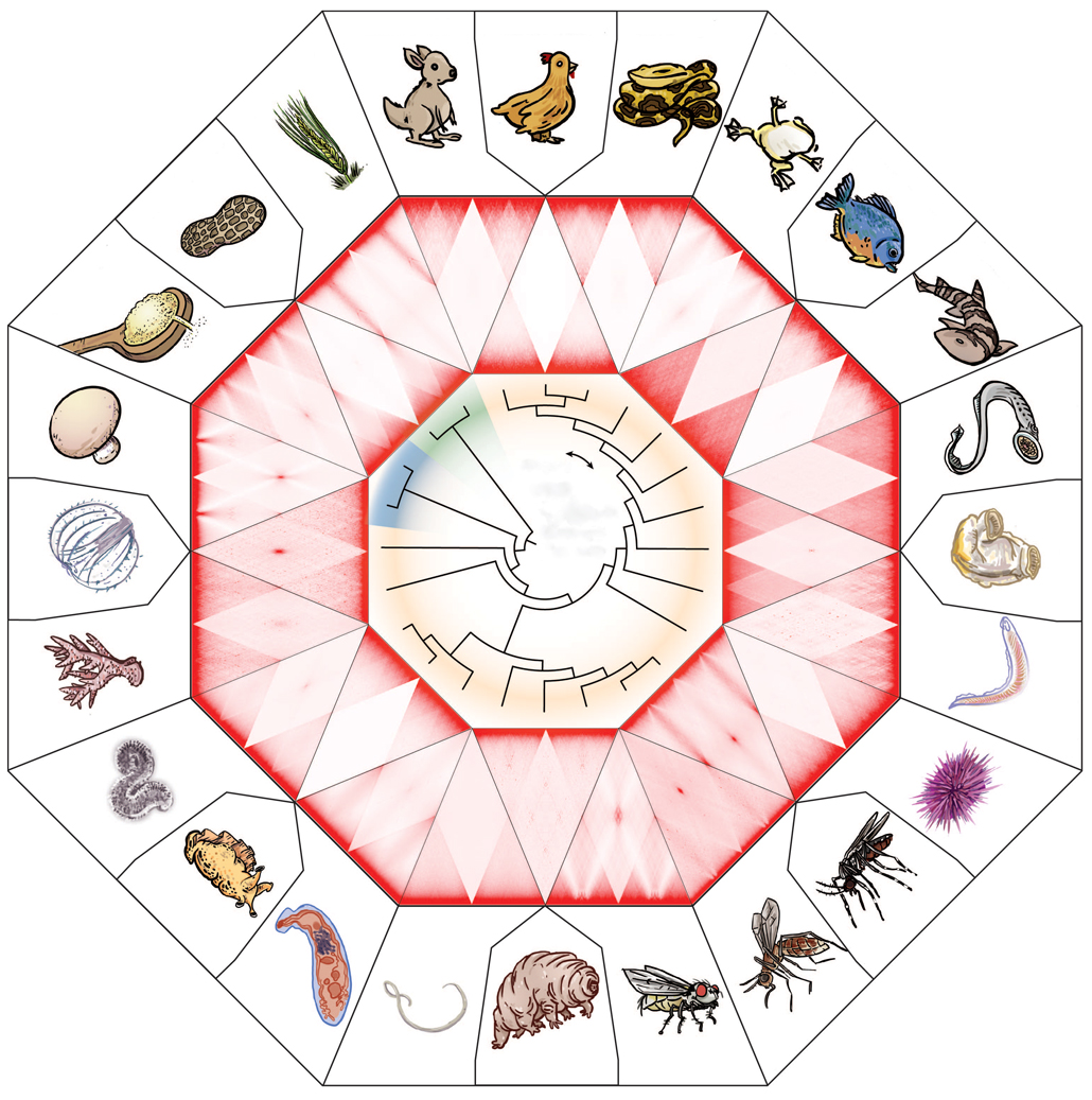
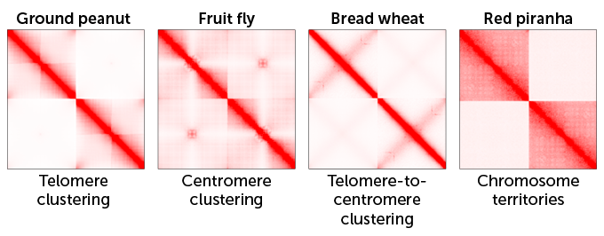
In the new study, Hoencamp and colleagues created heat maps that analyzed how chromosomes in the nuclei of 24 animal, plant and fungal species interacted inside their respective cells. The maps show the average number of connections among chromosomes in a cell’s nucleus — revealing how the genetic molecules fold — “on the scale from white to red,” says Olga Dudchenko, a genomicist at Baylor College of Medicine in Houston. “The more red, the more interactions. The less red, the less interactions.”
Chromosome contacts
A wide range of animal (colored yellow on the evolutionary tree at center), plant (green) and fungal (blue) species package chromosomes inside the cell nucleus one of two ways, based on how the chromosomes interact inside a nucleus. Heat maps here show the average number of interactions among chromosomes (darker shades of red indicate more interactions). Maps that have large, dark red triangles (such as for the red piranha, top right) indicate chromosomes that crumple up like a wad of paper. Other more checkerboard patterns (such as for the ground peanut, top left) give rise to chromosomes that fold more neatly, like stacked sheets of paper.
Patterns of chromosome interactions for 24 plants and animals
Adam Fotos, Olga Dudchenko, Benjamin Rowland and Erez Lieberman Aiden/Baylor College of Medicine
Throughout evolutionary history, organisms across the tree of life have switched among different packing methods, the researchers found. “We worked with a zoo of species, and [at first] it looked like a zoo of patterns of genome folding,” Dudchenko says. “Some maps would look like a checkerboard pattern. Other ones would look like a mattress with weird x’s.” Over time, it became clear that many of the same chromosome folding features were popping up again and again in different species.
Three types of interactions result in stacked sheets of chromosomes, giving the heat maps that checkerboard or mattress look. In one interaction, seen in the ground peanut (Arachis hypogaea) for example, the ends of different chromosomes tend to touch. In another, chromosomes from organisms like fruit flies touch in the middle. And in an interaction seen in bread wheat (Triticum aestivum), the arms of different chromosomes fold on top of one another.
Crumpled ball-like chromosomes, like those of the red piranha (Pygocentrus nattereri), sport a fourth type of interaction. In those structures, a chromosome folds in on itself in a tangle rather than touching other chromosomes, resulting in large red squares on the heat maps.
DNA dealings
Chromosomes interact in four different ways, resulting in two types of folding patterns. These selected heat maps for four species show the average number of interactions between two chromosomes; darker shades of red indicate more interactions. Three types of interactions — telomere clustering (ends of the two chromosomes touch), centromere clustering (middles touch) and telomere-to-centromere clustering (arms fold over each other) — make chromosomes fold like flat sheets of stacked paper. Interactions dubbed chromosome territories make chromosomes appear as crumpled balls: In this case, each chromosome is more likely to fold in on itself in a tangle, giving the appearance in the heat map of large, red squares.
C. Hoencamp et al/Science 2021
C. Hoencamp et al/Science 2021
Breaking parts of condensin II — a complex of proteins that helps assemble chromosomes as cells divide — can switch which organization a nucleus has. Tweaks to condensin II can make a crumpled human nucleus look like a folded fly’s nucleus, the team found. But some organisms have stacked sheets despite having intact condensin II. That means there may be other factors researchers haven’t yet found that push cells to cram chromosomes into the nucleus in a specific way, Hoencamp says.
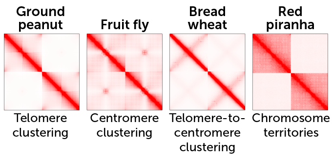
In the beginning, no one really understood how babies were made. Thinkers puzzled for millennia about how life arose from one generation to the next. But not until the 17th century did scientists start to seriously study the question. At that time, the theory of preformation held that minuscule humans already existed, fully formed, in either the mother’s menstrual blood or the father’s semen, depending on whether you were an “ovist” or a “spermist.”
Little changed until two late-19th century scientists, Oskar Hertwig from Germany and Hermann Fol from France, independently conducted experiments on sea urchins, proving conclusively that creating new offspring takes one egg and one sperm.
Despite the early confusion, the ancients were sure about one thing: Reproduction is far from a sure bet. Today, an estimated 15 percent of couples worldwide are unable to conceive a child naturally, leading to feelings of sorrow, loss and a profound sense of inadequacy for many. A century ago, science didn’t have much to offer these couples.
People used to think preformed humans grew inside sperm, as in this 1695 drawing.Nicolas Hartsoecker/Wikimedia Commons
The only fertility intervention widely available in 1921 was artificial insemination by donor sperm, which was morally and legally fraught. In the first half of the 20th century, the practice was often considered a form of adultery; as recently as 1963, an Illinois court ruled that a baby conceived this way, even with the husband’s consent, was illegitimate.
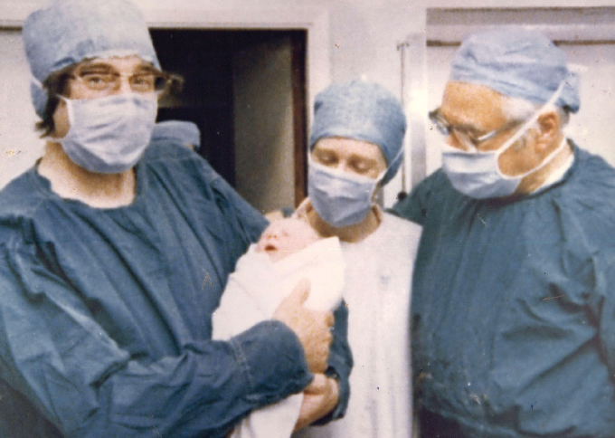
In 1978, everything changed. The birth of Louise Brown, the world’s first “test tube baby,” proved that infertile couples had another option: in vitro fertilization. The technique involved removing a mature egg from the mother, mixing it in a lab dish with the father’s sperm, and letting the fertilized egg, called a zygote, grow for a couple of days. The zygote was then returned to the mother’s uterus, where it could implant and grow in an otherwise normal pregnancy.
Since Brown’s historic birth, scientists have devised a range of ways to give Mother Nature a boost in baby-making. The various methods are known collectively as assisted reproductive technology, or ART. Some 9 million babies worldwide have been born using versions of ART.
The impact has been as profound sociologically as it has been medically. Now that ART has become almost routine, many of the early complaints about scientists playing God and manipulating life have faded away. Parenthood is now possible for people who never imagined it in their futures, including same-sex couples and single parents, thanks to such refinements as egg donors, surrogacy and the successful freezing of eggs, sperm and embryos. And all of it begins, as human life itself does, with the egg.
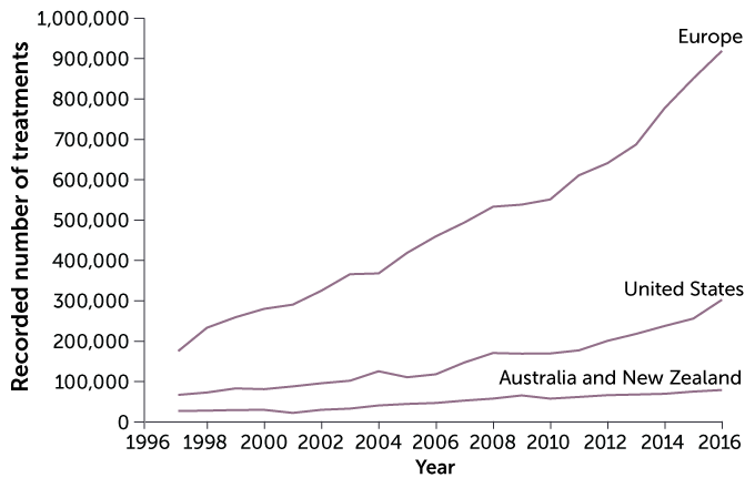
Helping hand
Assisted reproductive technology, or ART, offers several approaches to help people have a child. ART techniques are most popular in Europe.
Assisted reproductive technology procedures per year, 1997–2016
E. Otwell
E. Otwell
Source: C. De Geyter et al/Human Reproduction 2020
Good eggs
Even scientists can’t make babies without eggs. Normally, a woman produces only one mature egg every month, and the quality of her eggs tends to decline as she reaches her late 30s. So researchers’ ability to retrieve and prepare this scarce resource for fertilization, and if necessary preserve the eggs through freezing, have all been crucial in assisting reproduction.
Women have two ovaries, each one containing thousands of immature egg follicles. During the childbearing years, the ovaries usually release mature eggs in rotation: a single mature egg bursting from the right-hand ovary one menstrual cycle, from the left-hand ovary the next.
But women using ART often rely on injections of various fertility hormones to get the process going. These shots will enable sluggish ovaries to produce eggs that can be fertilized either through intercourse or in the lab via IVF. For IVF, the most robust-looking candidates are chosen to implant or to freeze for later use. This first step in ART, it turns out, is also one of the trickiest: choosing the right hormones to get the eggs you need.
This article is an excerpt from a series celebrating some of the biggest advances in science over the last century. For an expanded version of the story of human reproduction, and to see the rest of the series, visit Century of Science: The mystery of reproduction.
Knowledge about hormones and how they affect ovulation dates back to 1923, when scientists Edgar Allen and Edward Doisy of Washington University School of Medicine in St. Louis first isolated estrogen in experimental mice and rats and found that it was produced in the ovaries. By the 1940s, scientists had elucidated the ebbs and flows of other hormones in lab animals and humans — follicle stimulating hormone, luteinizing hormone and human chorionic gonadotropin — over the course of a typical menstrual cycle.
ART usually begins with a woman giving herself daily injections containing a cocktail of these hormones, generally for 10 to 14 days. But for some women who want to have children but are dealing with a cancer diagnosis, hormone injections aren’t an option — and the clock is ticking. They need to start cancer treatments as soon as possible, but many of the treatments are likely to damage the reproductive system.
To preserve their fertility, these women might choose to freeze their eggs before cancer treatment. But they might not have 10 or so days to wait until the hormone injections provoke their ovaries to produce extra eggs — nor might they be able to take the drugs in the first place if they have a hormone-sensitive cancer, such as some breast cancers, that ovulation-stimulating drugs might make worse. So for these women, researchers had to find ways to bring a bunch of eggs to maturity all at once and outside the body — a technique known as in vitro maturation.
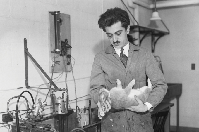
In vitro maturation was first used in 1934, when Harvard researchers Gregory Pincus and E.V. Enzmann used it in rabbits (SN: 3/10/34, p. 149). The two scientists cultured immature rabbit eggs for about a day, supplementing the nutrient broth either with extracts from cow pituitary glands or with an unspecified “maturity hormone.” Both supplements helped the immature eggs grow to maturity, at which point they were successfully fertilized.
In 1940, Pincus was asked by a New York Times reporter what the next big development might be in reproductive science. “There are no big steps, there are all little steps,” he said, declining to make any predictions. All he knew for sure, he said, was that the “big questions” of the day were: Why does an egg start to develop, and why does it continue to develop?
In the 1930s, Harvard researchers Gregory Pincus (shown) and E.V. Enzmann grew rabbit eggs to maturity and fertilized them in the lab. Pincus later went on to codevelop the birth control pill.Bettmann/Getty Images
When reproductive endocrinologists retrieve eggs from their ART patients, after either stimulating the ovaries via hormone injections or maturing the eggs in the lab, they have two choices: fertilize the eggs and implant the embryo right away, or store them. For women who are not yet ready to have a baby, storing the eggs is the best option. This is done through freezing — which, in the early days of ART, was a tricky business indeed. Eggs have a high fluid content that leads them to form crystals when frozen; during thawing, those crystals can damage the egg, especially the delicate apparatus needed to cut the cell’s chromosome number in half. By dividing the chromosomes, one egg plus one sperm can fuse together without doubling the chromosome count.
In the 1980s, egg freezing worked occasionally; the first successful pregnancy using a woman’s own frozen eggs, leading to the birth of healthy twins, was reported in 1986 by Christopher Chen of Flinders University of South Australia in Adelaide. But egg freezing was still a long shot. Estimates were that no more than 1 or 2 percent of thawed eggs would result in a live birth.
Then, in 1999, reports appeared of a more reliable freezing method: vitrification, which freezes the egg so rapidly that no ice crystals can form. A research team based in Australia and Italy described animal experiments in which 1 in 4 vitrified cow eggs were fertilized and later grew, by around day 5, to the blastocyst stage. It was only about half the rate achieved for fresh cow eggs, but it was still several times better than the rate for slow-frozen eggs. When it came to clinical use, some researchers put the live birth rate of vitrified eggs at about 2 to 12 percent for women under age 38.
At first, vitrification was limited to people who froze their eggs for medical reasons such as cancer. But in 2013, egg cryopreservation became an option for anyone who wanted to delay childbearing for any reason, medical or not.
By 2020, estimates were that a growing subset of women who choose egg vitrification each year in the United States do so because they’re not ready to have children yet but hope to eventually — lifestyle-related reasons that have come to be known as “social freezing.”
While social freezing is often promoted as a way to delay childbearing almost indefinitely, it turns out that most women never return to the clinic to use their frozen eggs. At McGill University in Montreal, for instance, William Buckett and his colleagues found that over the course of 13 years, the school’s cancer fertility preservation program treated 353 women, of whom 9 percent died, 6 percent got pregnant spontaneously, and the majority were either still dealing with their cancer or had lost touch with the clinic for unknown reasons. Just 23 women, 6.5 percent of the group, came back to McGill to use their frozen eggs or embryos. That low return rate is also true for women who opt for social freezing.
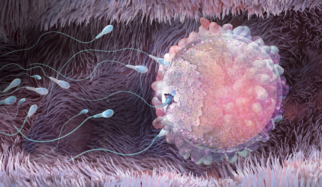
Once a single sperm has entered an egg, a special protein layer called the zona pellucida shuts out all other sperm.Nicolle Rager Fuller
Sperm meets egg
Compared with the human egg, sperm are pretty simple. They were first observed in 1677 when Antonie van Leeuwenhoek, the Dutch inventor of one of the world’s first microscopes, took a look at his own ejaculate under magnification and noticed what he called “animalcules” swimming around in the sample. But their structure and function didn’t come into focus until 1876, when Hertwig watched a sperm fertilize the egg of a sea urchin.
Pincus and Enzmann, who were the first to bring mammalian eggs to maturity in the lab, used rabbit sperm to achieve the first laboratory fertilization in a mammal in 1934.
It took years of struggle to make the leap from rabbits to humans. In 1951, an oddity of sperm made it seem that Pincus and Enzmann had gotten lucky. Sperm cells, it appeared, needed to be primed in some way through a process called capacitation before they could pierce the egg.
Robert Edwards of Cambridge University, one of the world’s leading investigators in IVF through the 1960s and 1970s, thought capacitation would be “a terrible obstacle to IVF,” recalls Roger Gosden, an embryologist who worked in Edwards’ lab and authored Edwards’ biography, Let There Be Life. He remembers some frantic attempts to mimic sperm capacitation — such as when scientists created porous chambers, roughly the size of an implanted IUD, or intrauterine device, used for contraception. The researchers would fill a chamber with sperm and insert it into the uterus of a volunteer, hoping to expose the sperm to whatever unknown capacitating substance exists in nature. After waiting a bit, the scientists would pull on a string attached to the chamber to retrieve the now “primed” sperm to see if they had indeed become better able to fertilize eggs in the lab.
In the end, scientists found an easier way. “You just have to wash the sperm to get rid of some surface constituents,” Gosden says, and the sperm are ready to fertilize an egg in a lab dish. Other difficulties in clinical research proved more formidable, such as gauging the best timing of egg retrieval from women, and tweaking how manbeatry days post-fertilization were best to transfer the zygote to the uterus. Edwards and his collaborators, including gynecologist Patrick Steptoe, had more than 300 failed attempts at in vitro fertilization before their first success with Louise Brown in 1978 in England (SN: 7/25/18). Edwards went on to win the Nobel Prize in 2010 for his IVF discoveries.
The 1978 birth of Louise Brown, a healthy baby girl, launched the field of in vitro fertilization. Although controversial at the time, IVF has been responsible for millions of births.ZUMA Press Inc./Alamy Stock Photo
Among the beneficiaries of Edwards’ pathbreaking work is Claudy, who was diagnosed with breast cancer at age 29, and who in a different century might never have been able to have a baby of her own. Claudy sought out Michaël Grynberg and his colleagues at the Antoine Béclère University Hospital fertility clinic, just outside Paris, to discuss ways to preserve her fertility. It was 2014, and egg freezing through vitrification was becoming more common. But bringing an immature egg to maturity in the lab was still relatively rare. From the time of the first baby born from fresh lab-matured eggs in 1991 to the time of Claudy’s arrival at Grynberg’s clinic, only about 5,000 such births had occurred.
But Grynberg had no choice. He had to retrieve immature eggs from Claudy, for the sake of both speed and to avoid aggravating her hormone-sensitive breast cancer with fertility drugs. In addition, he would have to do something unprecedented in the context of cancer: Freeze those lab-matured eggs for later use. No baby had ever been born from eggs taken from a cancer patient that were matured in the lab and then frozen. (There had been one baby born at McGill in 2009 from a woman who did not have cancer, whose eggs were matured in the lab and frozen, then thawed.)
Grynberg extracted seven immature eggs and was able to grow six of them to maturity over the next 48 hours. Those six went into the deep freeze, while Claudy had surgery and chemotherapy.
A few years later, Claudy’s oncologist told her it was safe to get pregnant, and she spent a year trying to conceive. But she didn’t. So in 2018, she returned to Grynberg’s clinic, where the doctors prepared to thaw her six frozen eggs.
To fertilize them, in Claudy’s case, required an additional bit of ART. Because her eggs had been frozen, her partner’s sperm would need help to fertilize her eggs. Vitrification causes changes in the egg’s outer membrane that makes the thawed egg particularly hard for sperm cells to penetrate. This membrane, called the zona pellucida, is a formidable barrier even in nature (SN: 1/3/09, p. 15). One of the first to describe it was Sardul Singh Guraya, a biologist at Punjab Agricultural University in India, who did his early work in field rats.
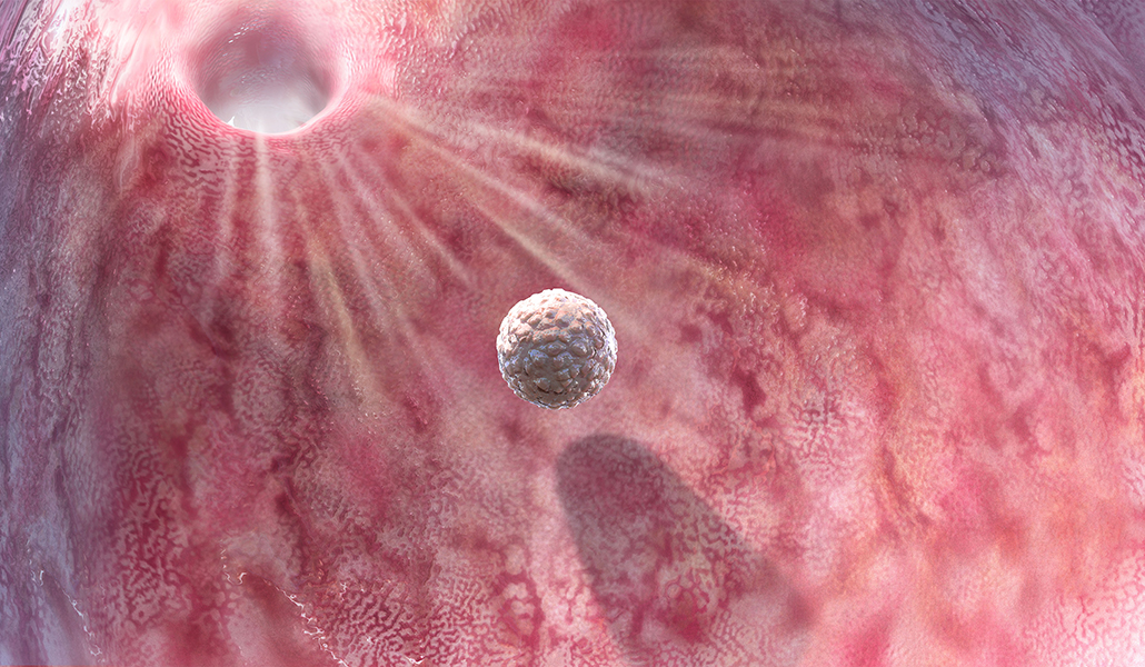
After fertilization, the blastocyst makes its way to the uterus. When an egg is fertilized in the lab, scientists can snip a cell from this developing mass to check for genetic problems without causing injury.Nicolle Rager Fuller
The zona pellucida, Guraya reported in 1978, is a barrier around the egg made of proteins and carbohydrates, and when one sperm breaches it, cortical granules rearrange themselves to shut out all other sperm. This ensures that the zygote will have a normal genetic complement of just two pairs of 23 chromosomes, one from the mother and one from the father, rather than a grossly inflated number that would result if multiple sperm fertilized the egg.
Scientists spent much of the next decade trying to get sperm into eggs that had been frozen using micro-manipulations described with such invasive terms as “zona drilling.” But the sperm still failed to reach the nucleus for fertilization.
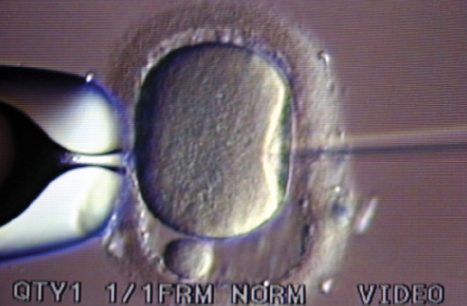
Then in 1992, Gianpiero Palermo, an Italian scientist on sabbatical from the University of Bari, reported on an accidental discovery he made while working in a fertility lab at the Free University of Brussels. When he tried to gently inject sperm beneath the outer layer of the egg, careful not to pierce the jellylike center known as the cytoplasm, he noticed that an occasional “dimple” in the membrane would allow the sperm to penetrate directly into the center anyway. When that happened, the egg was almost always fertilized. So despite the general recommendation to avoid doing so, Palermo tried injecting the sperm, tail and all, directly into the cytoplasm.
Of the first 47 attempts Palermo and his colleagues in Brussels made with this approach, 38 eggs remained intact after the injection, 31 were fertilized and 15 grew to embryos that could be transferred to a uterus.
While a pipette (left) holds an egg in place, a single sperm is injected to fertilize the egg in a technique known as ICSI.Hardas/Science Source
Ultimately, four babies were born: two healthy boys from two singleton pregnancies, and a healthy pair of boy-and-girl twins. The Belgian scientists called the procedure ICSI (pronounced ICK-see), shorthand for intracytoplasmic sperm injection.
Today, injecting a single sperm directly into an egg is even more common than the traditional form of IVF that adds sperm to an egg in a lab dish to let fertilization happen on its own. The injection method is used in about two-thirds of ART cycles around the world. And it is used in virtually all cycles that, like Claudy’s, start out with a hard-shelled frozen egg.
Growing a healthy baby
In 2018, Claudy returns to the fertility clinic at the Antoine Béclère University Hospital in suburban Paris. She is 34 years old and cancer-free. Because of the unusual nature of her case — her eggs were immature when they were retrieved and were brought to maturity in the laboratory — her doctors are not confident the eggs will survive the thawing and subsequent manipulations.
All six of Claudy’s eggs defrost with no apparent damage. The scientists perform ICSI on the eggs, using fresh sperm from Claudy’s partner. Five of the eggs fertilize.
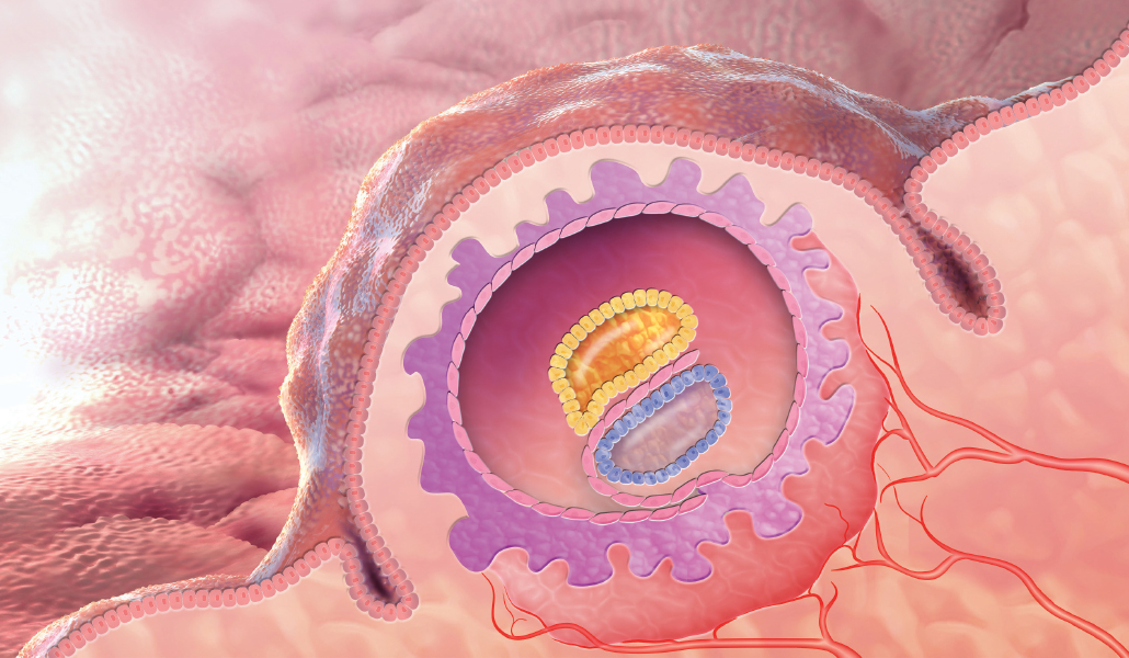
These five zygotes go into an incubator so they can develop to a stage that is ready to implant. They undergo the early stages of cleavage, in which one cell becomes two, two become four, four become eight, and so on.
At many fertility clinics elsewhere in the world, doctors might interrupt things at this point to snip off a cell or two from the early embryo to see if things are progressing normally. It was revolutionary to discover that this could even be done — a feat first accomplished in 1968 by embryologist Richard Gardner. At the time, Gardner was a graduate student working in Edwards’ lab in Cambridge. His work showed for the first time that it was possible in rabbits to take cells from a blastocyst without causing harm (SN: 8/3/68, p. 119).
Once implanted in the uterine wall, the embryo’s cells begin to differentiate, eventually forming all the tissues and organs that make up a human being.Nicolle Rager Fuller
Scientists can examine the chromosomes of those cells removed from a human embryo, a process called preimplantation genetic diagnosis, or PGD. They might be looking for a particular disease-linked gene that runs in the family, to avoid implanting an affected embryo in the uterus. Or they might be checking that a developing embryo has the right number of chromosomes, and that the embryo has a good chance of implanting in the uterus and coming out a baby with 10 fingers, 10 toes and the chance of a healthy life.
In the future, they might also use PGD to see whether a desired gene tweak, introduced via a gene-editing technique like CRISPR, has actually taken hold. Without PGD, none of these approaches, from disease prevention to designer babies, could take place.
Two days after putting Claudy’s five embryos in the incubator, only one is still undergoing cleavage. That is the embryo the doctors transfer to Claudy’s uterus in the autumn of 2018.
The embryo implants, and continues to develop the way any embryo would, no matter what its origin story — a ball of a few hundred genetically identical embryonic cells that eventually differentiate into the 200 or so cell types that make up a human being. The mechanism by which this occurs was first laid out in 1924 by the German investigator Hans Spemann, who discovered the “organizer effect” that leads particular regions of the embryo to develop into particular cell types.
In 1965, Beatrice Mintz created mice that wore their bizarre genetic lineage on their unique, black-and-white striped coats. In her lab at the Institute for Cancer Research in Philadelphia, she created a mouse with four parents — two mothers and two fathers — to demonstrate which parent’s genetic contribution ended up in which region of the body (SN: 4/12/69, p. 361).
Biologist Beatrice Mintz created thousands of four-parent mice, mixing different traits so she could trace the genetic origins of the distinctive organs that develop from the undifferentiated cells of a developing fetus.Smithsonian Institution/Flickr
Mintz merged eight-celled embryos from two different mice, one pure black embryo and one pure white, by putting them into a lab dish, dissolving the protective layer around each embryo, and actually smooshing them together using a glass rod. The result was a mosaic mouse: Some of its cells contained genes that could be traced directly to the two white mouse parents, and some had genes from the two black mouse parents.
Other mysteries of how embryos develop were revealed by “knockout” technology, in which scientists disabled genes in a particular region of an embryo to see what those genes controlled. In 1995, developmental biologists William Shawlot and Richard Behringer of the University of Texas MD Anderson Cancer Center in Houston reported using this method in mouse embryos, confirming Spemann’s theory that a tiny region of the embryo touches off changes in neighboring cells to turn them into particular cell types (SN: 4/1/95, p. 197).
The embryo in Claudy’s uterus develops normally; everything about her pregnancy seems ordinary — except how miraculous it might feel to Claudy herself, who must have had some doubt, as a young breast cancer patient, about whether she’d ever have a baby of her own. In early July 2019, Claudy goes back to the Antoine Béclère University Hospital, this time to give birth. Her son is born on July 6; she and her partner name him Jules.
When Grynberg asks Claudy’s permission to write up her landmark case in the Annals of Oncology, she is overwhelmed. “I thought about everything I had gone through,” she told a reporter for the British newspaper the Telegraph as she posed for a photo with baby Jules. “And I cried as I realized how lucky I was.”
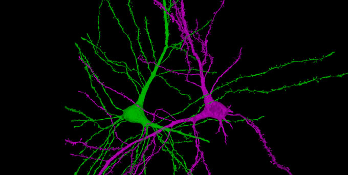
A new view of the human brain shows its cellular residents in all their wild and weird glory. The map, drawn from a tiny piece of a woman’s brain, charts the varied shapes of 50,000 cells and 130 million connections between them.
“It’s absolutely beautiful,” says neuroscientist Clay Reid at the Allen Institute for Brain Science in Seattle. “In the best possible way, it’s the beginning of something very exciting.”
Scientists at Harvard University, Google and elsewhere prepared and analyzed the brain tissue sample. Smaller than a sesame seed, the bit of brain was about a millionth of an entire brain’s volume. It came from the cortex — the brain’s outer layer responsible for complex thought — of a 45-year-old woman undergoing surgery for epilepsy. After it was removed, the brain sample was quickly preserved and stained with heavy metals that revealed cellular structures. The sample was then sliced into more than 5,000 wafer-thin pieces and imaged with powerful electron microscopes.
Computational programs stitched the resulting images back together and artificial intelligence programs helped scientists analyze them. A short description of the resulting view was published as a preprint May 30 to bioRxiv.org. The full dataset is freely available online.
These two neurons are mirror symmetrical. It’s unclear why these cells take these shapes. Lichtman Lab/Harvard University, Connectomics Team/Google
For now, researchers are just beginning to see what’s there. “We have really just dipped our toe into this dataset,” says study coauthor Jeff Lichtman, a developmental neurobiologist at Harvard University. Lichtman compares the brain map to Google Earth: “There are gems in there to find, but no one can say they’ve looked at the whole thing.”
But already, some “fantastically interesting” sights have appeared, Lichtman says. “When you have large datasets, suddenly these odd things, these weird things, these rare things start to stand out.”
One such curiosity concerns synapses, connection spots where signals move between nerve cells. Usually, most message-sending axons touch a message-receiving dendrite just once. In the new dataset, about 90 percent of the connections were these one-hit contacts. Some pairs of cells have slightly more contacts. But every so often, researchers spotted cells that connect multiple times, including one pair that were linked by a whopping 19 synapses.
Multiple connections have been spotted in mouse brains, though not quite as abundantly as in this human sample. And fly brains can also have many connections between cells, though they’re more dispersed than the newly described human connections, says neuroscientist Pat Rivlin of Howard Hughes Medical Institute’s Janelia Research Campus in Ashburn, Va. There, Rivlin works on the FlyEM Project, which aims to create detailed maps of the fruit fly nervous system.
The large dataset on the human brain provides a breakdown of just how common these types of connections are, says Reid. And that raises the question of what these extraordinarily strong synapses might be doing in the brain.
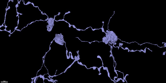
In their new database of human brain tissue, researchers found examples of unusual whorled nerve cell tendrils (purple), coiled like snakes.Lichtman Lab/Harvard University, Connectomics Team/Google
These cells might be able to compel their target cell into action in a powerful way, Lichtman speculates. Perhaps rote information, such as knowing 5 times 5 is 25 or knowing to stop at a red light, relies on these powerful inputs that efficiently drive information through the brain.
Molecular neuroscientist Seth Grant at the University of Edinburgh points out that although the map is a valuable tool, it shows only the anatomy of the brain. Other research will help clarify the function and composition of molecules that drive brain behavior. For now, the map is “very much an exploratory tool,” he says.
One curiosity to explore further is the team’s observation of two nerve cells, or neurons, that appeared to be entwined in a symmetrical dance. The images also revealed message-sending axons of neurons forming elaborate coils, unusual and mysterious whorls that look like coiled snakes. “We had just never seen anything like it,” Lichtman says. Once the researchers knew how to look for these coils, more and more turned up.
These extremely detailed brain maps are a culmination of years of research, says Reid, who is working on maps of mouse and human brains at the Allen Institute (SN: 8/7/19). “It’s this magical time in history” when the map-making tools, such as computational methods, machine learning and powerful microscopes, are all available, Reid says. “This work is just beginning to see the light of day.”
What these maps will ultimately reveal is still anybody’s guess. Lichtman is circumspect about whether these maps will lead to a deep understanding of the brain. “I think the best we can do is describe,” he says. “I hope that at some point, we will get to a place where we are no longer surprised by what we see.”


 Researchers at UC San Diego, San Diego State University, and international collaborators have designed and validated a prediction model to signal counties at risk of future overdose death outbreaks.